
A Dental X-ray enables dentists to identify potential oral health issues at an early stage, allowing for accurate and timely treatment planning. In the article below, Shark Dental Clinic provides comprehensive information about modern dental X-rays types, costs, and important precautions—helping ensure your examination is safe, comfortable, and effective.
What are dental X-ray?
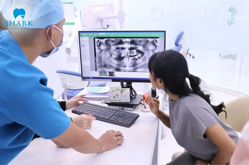
Dental X-rays are a diagnostic tool that utilizes X-rays—a form of high-energy radiation—to penetrate soft tissues and capture detailed images of the internal structures of the oral cavity. These images allow dentists to observe teeth, tooth roots, jawbone, dental pulp, and surrounding tissues that are not visible to the naked eye.
With this technology, dentists can accurately assess oral health, detect dental issues such as tooth decay, infections, abscesses, and impacted or misaligned wisdom teeth at an early stage, and monitor treatment outcomes following dental procedures.
How do dental radiographs work?
The operation of a dental X-ray is similar to that of X-ray imaging used for other parts of the body. The machine emits a small amount of radiation that passes through soft tissues like the lips, gums, and cheeks, capturing realistic images of the teeth and underlying jawbone structures.
Currently, there are 2 commonly used methods for dental X-rays, each with a different operating mechanism:
- Traditional X-ray: This method uses specialized film to capture images.
- Digital X-ray: This method employs electronic sensors connected to a computer, allowing images to be displayed immediately on the screen with high clarity.
Modern digital dental X-ray technology reduces radiation exposure by approximately 80–90% compared to traditional film-based methods. This not only accelerates the diagnostic process but also maximizes safety for patients.
What are the most common types of dental X-rays?
Dental X-rays are divided into two main categories: intraoral X-rays and extraoral X-rays. The type prescribed depends on the examination purpose and the patient’s oral condition.
Intraoral dental X-rays
Intraoral X-rays are the most common type, where the film or sensor is placed inside the patient’s mouth. This technique provides highly detailed images of the teeth, alveolar bone, and surrounding supporting tissues.
- Bitewing X-ray: Helps detect cavities between teeth and evaluates the condition of the supporting bone below the gum line.
- Periapical X-ray: Provides a complete image of the entire tooth, from the crown to the root tip, aiding in the diagnosis of pulp inflammation, bone loss, and periapical diseases.
- Occlusal X-ray: Commonly used to detect impacted teeth, misaligned teeth, or abnormalities within the oral cavity.
Extraoral dental X-rays
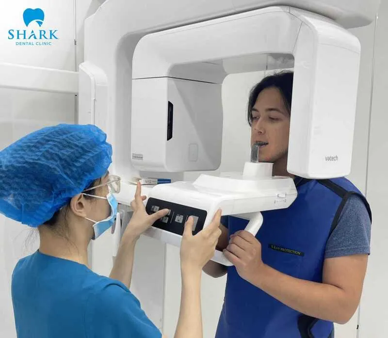
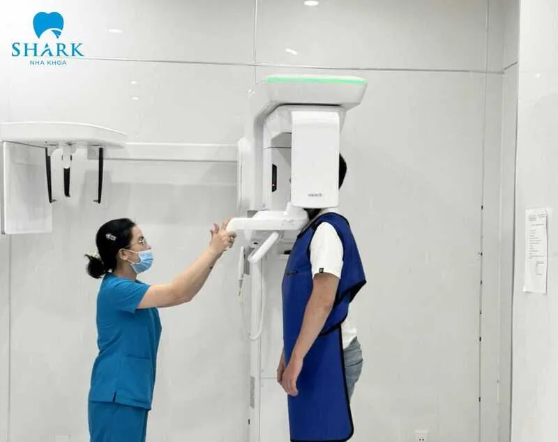
With extraoral X-rays, the film or sensor is placed outside the oral cavity, offering a comprehensive view of the entire face, jawbone system, and related joints.
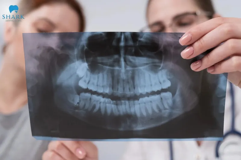
- Panoramic X-ray: Provides an overall image of the entire dentition, jawbone, nerves, and sinuses, effectively supporting diagnosis and comprehensive treatment planning.
- Cephalometric X-ray: Produces a lateral view of the head and is commonly used in orthodontics to analyze bite alignment and facial skeletal structure.
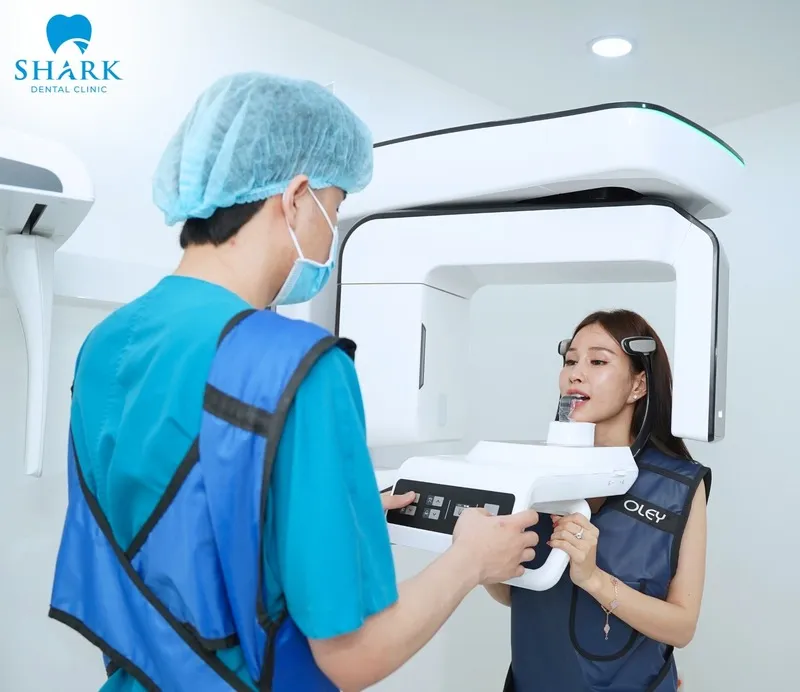
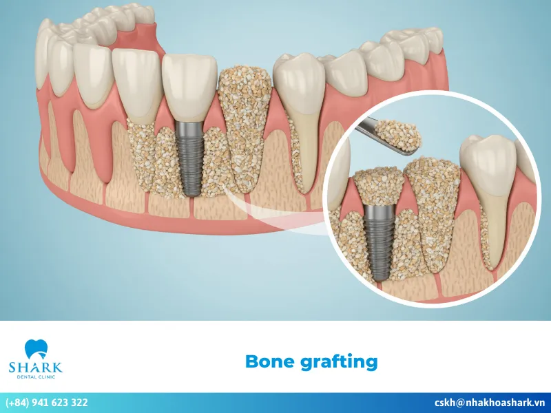
- Cone Beam CT: An advanced technology that delivers detailed 3D images of teeth, jawbone, nerves, and sinuses. It is often indicated for dental implant placement, surgery, and complex dental treatments.
How much is the cost of a dental X-ray?
The cost of a dental X-ray ranges from approximately 50,000 to 500,000 VND per scan, depending on the type of imaging. Here are the typical price ranges:
- Periapical dental X-ray: 50,000 – 100,000 VND per scan
- Panoramic dental X-ray: 150,000 – 250,000 VND per scan
- Cephalometric X-ray: 200,000 – 300,000 VND per scan
- Cone Beam CT Scan: 250,000 – 500,000 VND per scan
Please note that these prices are for reference only and can vary based on several factors, including the type of imaging film or sensor used, the number of images required, the technology applied, and the specific dental clinic. To prepare for your visit, it’s advisable to check the publicly listed price table on the dental clinic’s official website and choose a service that best fits your needs.
Are dental X-rays safe?
Dental X-rays are considered safe because they use a very low dose of radiation, which is strictly regulated according to medical safety standards. The amount of radiation exposure from dental imaging is comparable to the natural background radiation we encounter daily from our environment, building materials, or air travel at high altitudes.
However, frequent exposure to high doses of radiation may have adverse health effects. For this reason, dentists recommend dental X-rays only when necessary for diagnosis and treatment planning. Before each scan, dentists carefully assess the diagnostic benefits against safety considerations to ensure maximum protection for the patient’s health.
How often are dental X-rays needed?
The frequency of dental X-rays is not fixed and varies based on factors such as age and an individual’s oral health condition. Generally, dentists recommend periodic dental imaging to monitor oral health and detect potential issues early. Typical recommendations include:
- Adults: Dental X-rays every 2–3 years
- Adolescents: Recommended every 1.5–3 years
- Children: Dental X-rays every 1–2 years
For individuals with a history of tooth decay, gum disease, or a high risk of oral health problems, dentists may suggest more frequent imaging—typically every 6 months to 1.5 years for adults and every 6–12 months for children.
How long should you wait between dental X-rays?
The interval between dental X-rays is flexible, adjusted to ensure accurate diagnosis while maintaining patient safety. Depending on oral health conditions, dentists might recommend:
- For patients with stable oral health: An interval of 1–2 years is generally appropriate. This allows for early detection of subtle changes in tooth and jawbone structures.
- For patients undergoing treatment: If you are receiving root canal treatment, periodontal therapy, or dental implant placement, the interval may be shortened to every few months. This helps the dentist closely monitor healing progress and promptly address any complications.
Before and after considerations for the patient
Before and after undergoing a dental X-ray, patients should keep several important considerations in mind to ensure the procedure is safe, accurate, and provides optimal diagnostic results.
Before a dental X-ray:
- Inform your dentist about your health status: This is especially important if you are pregnant or suspect you might be pregnant, so the dentist can take necessary precautions or postpone the scan if it is not urgent.
- Remove metal objects: Take off removable dentures or metal accessories, as metal can block X-rays and create shadows that interfere with diagnostic accuracy.
- Stay relaxed: Dental X-ray procedures are painless. Simply maintain the correct position as instructed by the technician for a few seconds to avoid blurred images.
After the dental X-ray:
- Listen to your dentist’s consultation: Digital dental X-ray images are usually available immediately. Discussing the results with your dentist helps you understand your current oral condition and the next steps in your treatment plan.
- Resume normal activities: Dental X-rays do not cause side effects or fatigue. You can eat, work, and drive normally without any special rest or restrictions.

In summary, dental X-rays play a vital role in the early detection of oral health issues and assist dentists in making accurate treatment decisions. Understanding the types of imaging, costs, and important considerations can help ensure a safe and comfortable examination experience. For international-standard dental X-ray services, contact Shark Dental Clinic at (+84) 941 623 322 for prompt consultation and appointment scheduling.


Comment on the post